We're sorry. An error has occurred
Please cancel or retry.
Medical Imaging for the Health Care Provider

Some error occured while loading the Quick View. Please close the Quick View and try reloading the page.
Couldn't load pickup availability
- Format:
-
28 August 2023

AJN award winner!
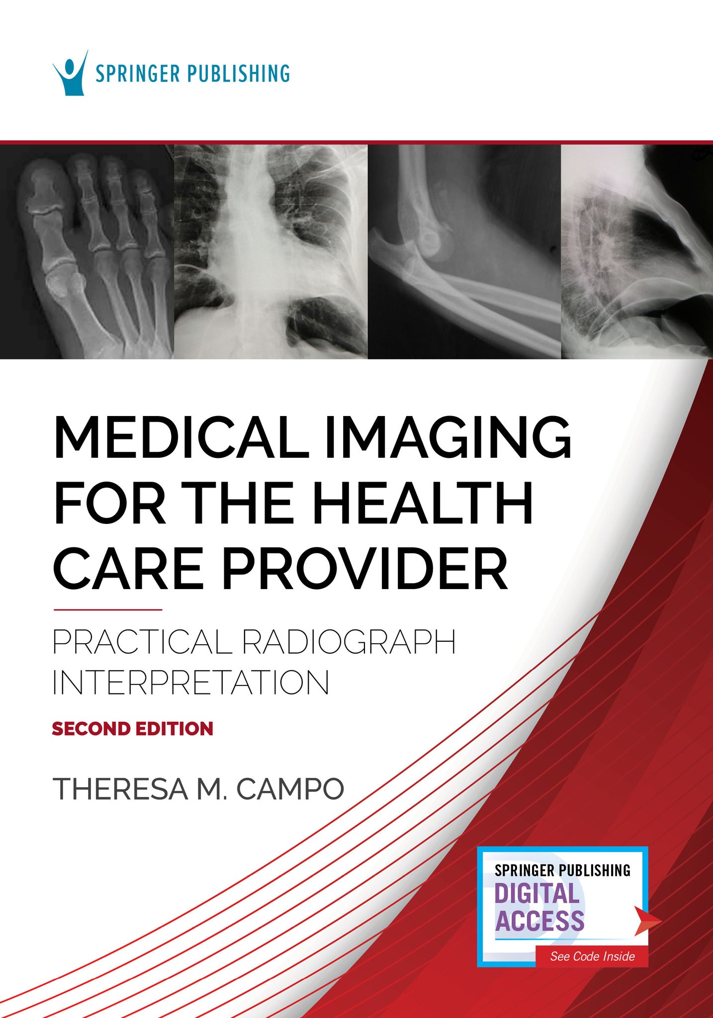
This is a concise, easy-to-use reference, enabling health care providers to identify and understand how and when to use the full scope of medical imaging testing modalities-- radiographs, CTs, nuclear imaging, and ultrasound scans and images. The new second edition features a more in-depth discussion of each modality with a focus on the foundational concepts of radiography interpretation of the chest, abdomen, extremities, and spine. It expands coverage of imaging and increases the number of images provided for a total of 400. In addition, the Springer Connect website includes dozens of videos to greatly enhance the learning process.
With clear descriptions of each modality—supported by figures, tables, and actual patient films—the text guides readers through the clinical decision-making process. It describes how to choose the best diagnostic test to assess a presenting condition, and examines interpretations of plain radiographs of the chest, abdomen, extremities, and spine. The book fosters an in-depth understanding of the differences between modalities, their attributes, and an appreciation for their parameters with age-appropriate considerations. To assist health care practitioners with the challenges of interpreting plain radiographs, the book simplifies this process with an incremental approach to correct interpretation of what appears on the radiograph and understanding the rationale behind the interpretation. Purchase includes online access via most mobile devices or computers.
New to the Second Edition:
- In-depth discussions of different medical imaging testing modality, with a focus on foundational concepts of radiology interpretation of the chest, abdomen, extremities, and spine
- Exploration of similarities and differences between modalities
- Over 400 images
- Accompanying videos available via Springer Connect
Key Features:
- Addresses the basics of radiology, CT scans, nuclear imaging, MRIs, and ultrasound and their characteristics and differences
- Provides a step-by-step approach to interpretation of radiographs
- Guides in the selection of the correct diagnostic test
- Supports information with figures, tables, images, and films
- Useful to a wide range of nurse practitioners, physician assistants, and other providers in multiple settings


Reviewers
Foreword by David Begleiter, MD
Preface
Acknowledgements
Springer Publishing ConnectTM Resources
SECTION I: INTRODUCTION TO MEDICAL IMAGING INCLUDING RADIOGRAPHS, CT, NUCLEAR SCANS, MRIS, AND ULTRASONOGRAPHY
Chapter 1. Radiology Basics, History of Radiology
- Factors Affecting Images
- Conclusion
- Resources
Chapter 2. Radiating Testing Modalities
- Radiographs
- Computer Tomography (CT)
- Nuclear Scanning
- Conclusion
- Resources
Chapter 3. Nonradiating Testing Modalities
- Magnetic Resonance Imaging (MRI)
- Ultrasonography
- Considerations When Ordering Diagnostic Medical Imaging
- Conclusion
- Resources
SECTION II: INTERPRETING CHEST AND ABDOMINAL RADIOGRAPHS
Chapter 4. Basic Interpretation of the Chest
- Radiographic Densities
- Adequacy of Radiographs
- Pediatric Considerations (Comparison of Adult and Infant/Child)
- Mediastinal Width
- Conclusion
- Resources
Chapter 5. Abnormalities Found on Radiographs of the Chest
- Atelectasis
- Pulmonary Edema
- Pleural Effusion
- Interpretation of Infiltrates and Consolidation
- Pneumothorax
- Tension Pneumothorax
- Pneumomediastinum
- Hyperaeration
- Masses and Tumors
- Conclusion
- Resources
Chapter 6. Basic Interpretation of the Abdomen
- Interpretation and Normal Findings
- Free Air and Air-Fluid Levels
- Calcifications
- Foreign Bodies
- Dilated Small Bowel
- Dilated Large Bowel and Megacolon
- Conclusion
- Resources
SECTION III: INTERPRETATION OF EXTREMITY RADIOGRAPHS
Chapter 7. Basic Interpretation of Long Bone--Upper Extremity Radiographs
- Normal
- Abnormalities of the Upper Extremity
- Bone Lesions
- Conclusion
- Resources
Chapter 8. Basic Interpretation of Long Bone--Lower Extremity Radiographs
- Normal
- Describing Fractures
- Pediatric Considerations
- Abnormalities of the Lower Extremity
- Conclusion
- Resources
SECTION IV: INTERPRETATION OF SPINE RADIOGRAPHS
Chapter 9. Basic Interpretation of Cervical Spine Radiographs
- Normal
- Abnormalities
- Conclusion
- Resources
Chapter 10. Basic Interpretation of Thoracic Spine Radiographs
- Normal
- Abnormalities
- Conclusion
- Resources
Chapter 11. Basic Interpretation of Lumbar Spine Radiographs
- Normal
- Abnormalities
- Conclusion
- Resources
Index


