


Some error occured while loading the Quick View. Please close the Quick View and try reloading the page.
The relentless pace of innovation in biomedical imaging has provided modern researchers with an unprecedented number of techniques and tools to choose from. While the development of new imaging techniques is vital for ongoing progress in the life sciences, it is challenging for researchers to keep pace. Imaging Modalities for Biological and Preclinical Research is designed to provide a comprehensive overview of currently available biological and preclinical imaging methods, including their benefits and limitations. Experts in the field guide the reader through both the physical principles and biomedical applications of each imaging modality, including description of typical setups and sample preparation.
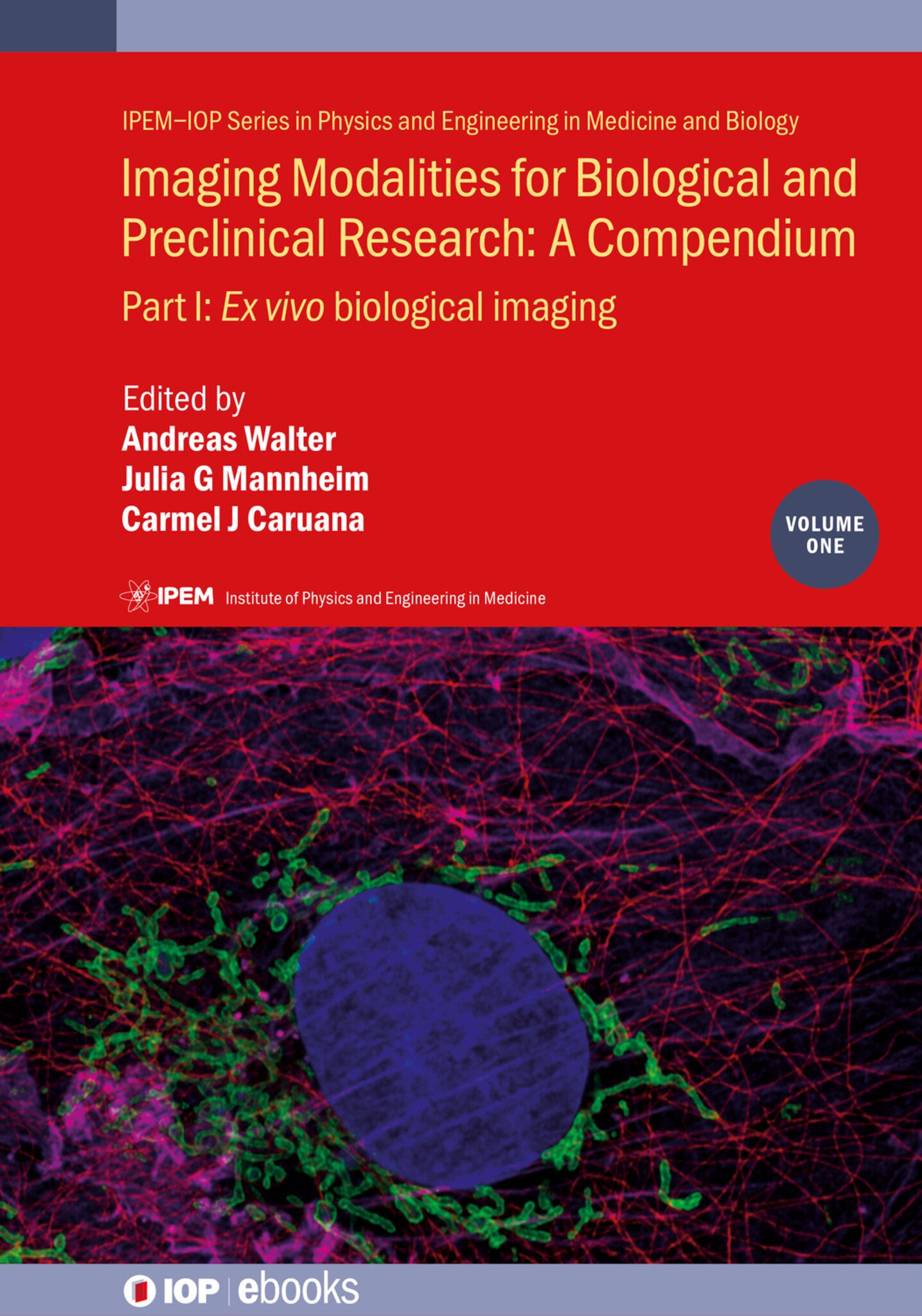
Volume 1 focuses on ex-vivo imaging. It covers all available advanced and basic light and fluorescence microscopy modalities, X-ray, electron, atomic force and helium ion microscopy, dynamic techniques such as fluorescence recovery after photobleaching as well as spectroscopic techniques such as coherent Raman imaging or mass spectrometry imaging.
Key features
- Provides an overview of fast-evolving ex-vivo imaging technologies.
- Bridges biological and preclinical imaging.
- Written by imaging specialists with extensive expertise in their respective fields.


Preface
Part I: Ex-vivo Imaging
I.1 - Light and fluorescence microscopy
I.1.a Transmission light microscopy
I.1.b Fluorescence and confocal microscopy
I.1.c Lensless digital inline holographic microscopy
I.1.d High-content microscopy
I.1.e Calcium imaging
I.1.f Fluorescence cryo-microscopy
I.2 - Light microscopy of tissues and thick samples
I.2.a Light sheet fluorescence microscopy (LSFM)
I.2.b Lattice light sheet microscopy
I.2.c Multiphoton microscopy
I.2.d Second and third harmonic generation imaging
I.2.e Adaptive optics
I.2.f Optical projection tomography
I.2.g High-resolution episcopic microscopy (HREM)
I.2.h Tissue image cytometry
I.2.i Histopathology
I.3 - Super-resolution microscopy
I.3.a Total internal reflection fluorescence microscopy
I.3.b Structured illumination microscopy
I.3.c Single-molecule localization microscopy (SMLM)
I.3.d Stimulated emission depletion (STED) microscopy
I.3.e Expansion microscopy
I.3.f Scattering-type scanning near-field optical microscopy (s-SNOM)
I.4 - X-ray microscopy I.4.a Hard x-ray tomographic microscopy
I.4.b Soft x-ray tomography
I.5 - Electron microscopy I.5.a Transmission electron microscopy
I.5.b Cryo-transmission electron microscopy
I.5.c Scanning electron microscopy
I.5.d Volume SEM
I.5.e Nanotomy
I.5.f Electron microscopy—STEM
I.6 - Atomic force microscopy and spectroscopy
I.7 - Helium ion microscopy
I.8 - Dynamic techniques I.8.a Förster resonance energy transfer (FRET)—fluorescence lifetime imaging microscopy (FLIM)
I.8.b Fluorescence correlation spectroscopy (FCS)
I.8.c Fluorescence recovery after photobleaching (FRAP)
I.8.d Single particle tracking
I.8.e BIOSPECKLE imaging
I.9 - Imaging and spectrometry I.9.a Raman imaging
I.9.b Coherent Raman imaging (CARS, SRS)
I.9.c Brillouin microscopy
I.9.d Chemical analysis—EELS and EFTEM
I.9.e Chemical analysis—EDX
I.9.f Micro-x-ray fluorescence analysis
I.9.g Mass spectrometry-based imaging
I.9.h Imaging mass cytometry
I.9.i Magnetic resonance microscopy
I.10 - Autoradiography