We're sorry. An error has occurred
Please cancel or retry.
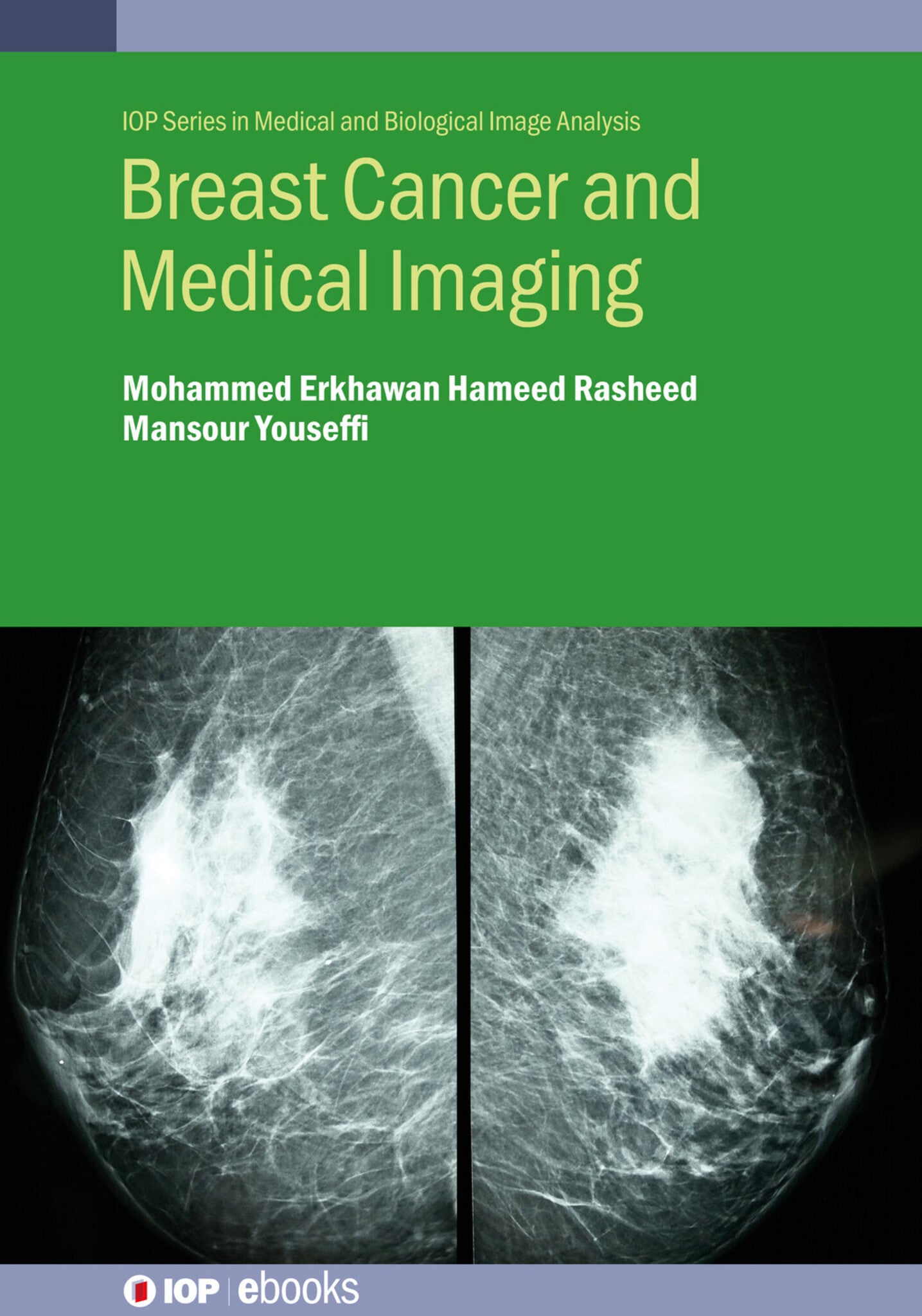
Breast Cancer and Medical Imaging

Some error occured while loading the Quick View. Please close the Quick View and try reloading the page.
Couldn't load pickup availability
- Format:
-
05 March 2024

Breast cancer is a disease that develops in the breast cells and progresses in stages. A few early symptoms may include a new lump in the underarm or in the breast, itching or discharge from the nipple, and skin texture change of the nipple or breast. Approximately 1.3 million people per year worldwide are diagnosed with this condition. As with any cancer, early detection is a key to less traumatic treatment and ultimately complete recovery.
Key Features:
-
Detailed in-depth coverage of breast cancer in all critical aspects such as prevention, detection, diagnosis, and treatment
-
Presents an overview of the relevant medical imaging modalities used for breast cancer
-
Discusses the pros and cons of each imaging modality
-
Book presents detailed breast cancer imaging clinical case studies

MEDICAL / Allied Health Services / Imaging Technologies, Pre-clinical medicine: basic sciences, MEDICAL / Oncology / General, Oncology, Surgical oncology

Preface
Acknowledgements
Author biographies
List of abbreviations
1 Introduction
References
2 Breast cancer stages and risk factors
2.1 Background
2.2 Breast cancer stages
2.2.1 Stage 0
2.2.2 Stage I
2.2.3 Stage II
2.2.4 Stage III
2.2.5 Stage IV
2.3 Breast cancer risk factors
2.3.1 Age
2.3.2 Family history and genetics
2.3.3 Taking oral contraceptives
2.3.4 Taking hormone replacement therapy (HRT)
2.3.5 Pregnancy history
2.3.6 Race and ethnicity
2.3.7 Breast composition (density)
2.3.8 Drinking alcohol and its effect on breast cancer
References
3 Breast cancer prevention and breast cancer types
3.1 Breast cancer prevention
3.1.1 Modification of lifestyle and eating habits
3.1.2 Early pregnancy and breast feeding
3.1.3 Taking medicines
3.1.4 Prevention through surgery
3.2 Breast cancer control
3.3 Breast cancer symptoms
3.4 Breast cancer types
3.4.1 Ductal carcinoma in situ (DCIS)
3.4.2 Invasive ductal carcinoma (IDC)
3.4.3 Lobular carcinoma in situ (LCIS)
3.4.4 Invasive lobular carcinoma (ILC)
3.4.5 Inflammatory breast cancer (IBC)
3.4.6 Paget’s disease of the nipple
3.4.7 HER2-positive breast cancer
3.5 Prevalence of breast cancer in young women
3.6 Breast cancer and pregnancy
3.7 Breast cancer in men
References
4 Breast cancer treatments
4.1 Surgery
4.2 Chemotherapy
4.3 Radiotherapy (RT)
4.4 Hormone treatment
4.5 Biological therapy (targeted therapy)
4.6 Complementary and alternative medicine (CAM)
4.6.1 Cannabidiol
4.6.2 Graviola (soursop)
4.6.3 Origanum vulgare
4.6.4 Zamzam water
4.6.5 Traditional Chinese Medicine (TCM)
4.6.6 Paris polyphylla
4.7 Breast cancer recurrence
4.8 Blood marker test
References
5 Medical imaging
5.1 Background
5.2 Ionising radiation medical imaging techniques
5.2.1 X-ray (conventional radiography)
5.2.2 Mammography (mammogram)
5.2.3 Computed tomography (CT)
References
6 Non-ionising radiation medical imaging techniques
6.1 Ultrasound imaging (USI)
6.2 Magnetic resonance imaging (MRI)
References
7 Radionuclide medical imaging techniques
7.1 Introduction
7.1.1 Positron emission tomography (PET)
7.1.2 Single-photon emission computed tomography (SPECT)
7.2 Medical image analysis
7.3 DICOM standard and Merge PACS
7.4 Medical image quality
7.5 Artificial intelligence (AI) and breast cancer imaging
References
8 Breast cancer and medical imaging clinical case studies
8.1 Information governance
8.2 Introduction to genetic counselling
8.3 Dense breast tissue case
8.3.1 Medical history of the patient
8.3.2 Breast imaging reporting and data system (BI-RADS)
8.3.3 Mammography of the patient
8.3.4 Ultrasound imaging of the patient
8.3.5 Treatment of the patient
8.3.6 Discussion
8.4 Metastatic breast cancer case
8.4.1 Breast screening mammography
8.4.2 Breast cancer and bone metastasis on a PET/CT scan of the patient
8.4.3 Discussion
8.5 High grade (G3) triple-negative metaplastic breast cancer
8.5.1 Introduction
8.5.2 Medical history of the patient
8.5.3 The patient’s family history of cancer
8.5.4 Diagnostic tests
8.5.5 Treatment
8.5.6 Discussion
References
9 Discussion and conclusion
9.1 Discussion
9.2 Conclusion
9.3 Recommendations
9.4 Implications
References


