


Some error occured while loading the Quick View. Please close the Quick View and try reloading the page.
Magnetic resonance imaging (MRI) is a scan that uses strong magnetic fields and radio waves to produce detailed images of the inside of the body.
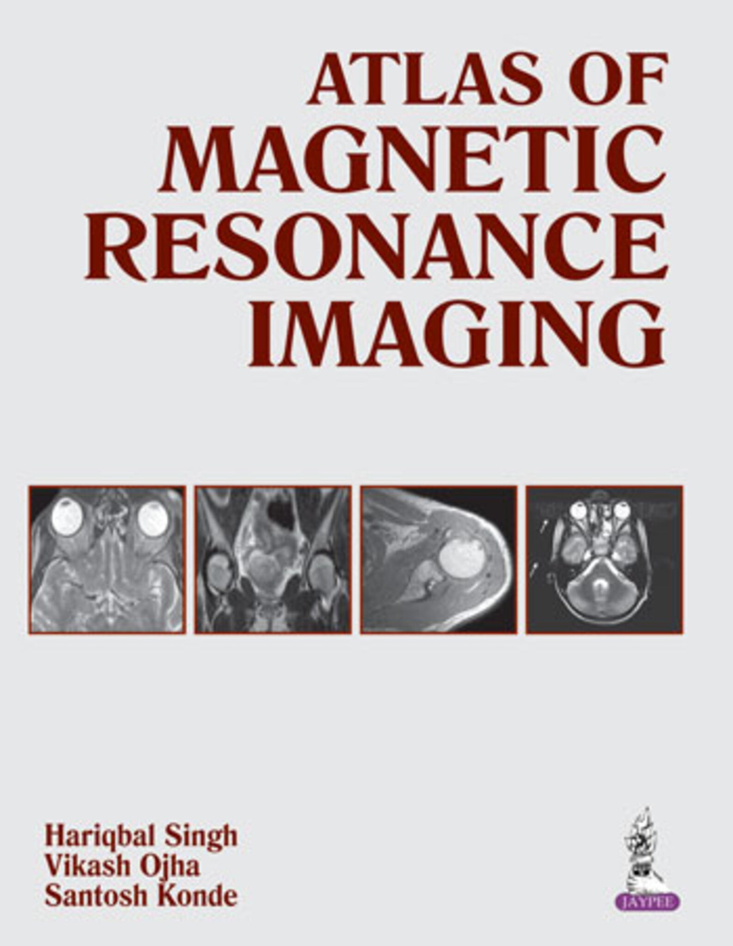
This atlas is a comprehensive guide to MRI for radiology trainees and practising clinicians. Beginning with an introduction to the technique and the associated physics, each of the following chapters presents numerous high quality MRI images of different body systems including brain, orbit, spine, pelvis, and musculoskeletal system. Images are accompanied by detailed descriptions and each topic begins with a section on relevant anatomy.
A self assessment chapter is included to test knowledge, and the final chapters include a glossary of MRI terms and MRI acronyms.
Key points
- Comprehensive guide to MRI for trainees and radiologists
- In depth coverage of different body systems
- Topics illustrated by high quality MRI images with descriptions
- Includes self assessment section


Hariqbal Singh MD DMRD
Professor and Head, Department of Radiology, Smt Kashibai Navale Medical College and General Hospital, Pune, Maharashtra, India
Vikash Ojha MD
Consultant, Department of Radiology, Apollo Jehangir Hospital, Pune, Maharashtra, India
Santosh Konde MD
Associate Professor, Department of Radiology, Smt Kashibai Navale Medical College and General Hospital, Pune, Maharashtra, India
- Physical Principle of Magnetic Resonance Imaging
- Brain
- Orbit
- Spine
- Pelvis
- Musculoskeletal System
- Miscellaneous
- Self-Assessment
- MRI Artifacts
- MRI Contrast
- Glossary of MRI Terms
- MRI Acronyms